
You can visualize the size and location of your own heart by following these simple steps.
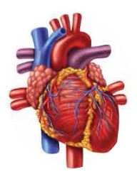
First, make a fist and place it in the middle of your chest, 2-3cm below the collar bone. Then, move the fist 2-3 cm slightly to the left and imagine it sitting in between your chest and back. This would be the location of your heart. Anatomically, the heart sits between the left and right lungs, behind the sternum and above the diaphragm. The heart is surround by a protective layer, the pericardium, that allows pumping activity of the heart without friction against the lungs and bones (The Cardiovascular system, 2000).
The heart and circulatory system (blood vessel system throughout the whole body) make up the cardiovascular system. This important system delivers oxygen and nutrients to every cell in the body, and removes carbon dioxide and waste products. Oxygen-rich blood is carried from the heart to the rest of the body through a complex network of arteries and capillaries. Oxygen-poor blood is carried back to the heart from the cells through the veins (The heart and Stroke Foundation, 2015).
The heart muscle, called the myocardium, is very unique; it is the only muscle in our body that never stops working. Usually, muscles work only when they are needed. For example, leg muscles work only when we stand or walk, whereas, the heart works constantly, even when we sleep. Another special feature of the heart muscle is that it works by electrical impulses. The heart has a receptor that analyzes blood pressure and oxygen level. Once the analysis is done, the heart sends electric signals to the muscles of the four chambers, which have to work accordingly to propel blood out of the heart (The Heart and Stroke Foundation, 2015). If there is a small impairment of electric transmission within the heart, the heart beat becomes irregular and even stops in severe cases.